Just In
- 1 hr ago

- 5 hrs ago

- 5 hrs ago

- 11 hrs ago

Don't Miss
- Movies
 LSD2 Box Office Collection Day 1 Prediction: Dibakar Banerjee’s Film To Have Slow Start; Will Fail To Touch 5C
 - Finance
 1:3 Bonus Share, Rs 13.25/Share Dividend: Buy Maharatna PSU, TP Rs 355, Fundraise Approved
 - News
 12 Jurors Picked For Donald Trump’s Hush Money Trial, Alternate Selection Continues
 - Sports
 Who Won Yesterday's IPL Match 33? PBKS vs MI, IPL 2024 on April 17: Mumbai Indians Escape Last-Ditched Fight by Punjab Kings To Win
 - Automobiles
 Aprilia RS 457 Accessories: A Detailed Look At The Prices
 - Education
 Karnataka SSLC Result 2024 Soon, Know How to Check Through Website, SMS and Digilocker
 - Technology
 Nothing Ear, Ear a With ANC, Up to 42.5 Hours of Battery Launched; Check Price and Availability
 - Travel
Telangana's Waterfall: A Serene Escape Into Nature's Marvels
Digital Myxoid Cyst: Symptoms, Causes And Treatment
A digital myxoid cyst is a benign and small lump or swelling that usually develops at the base of fingernails or toenails. These non-cancerous lumps are also termed as digital mucous cyst or mucous pseudocyst. A myxoid cyst is usually not painful and in most cases, does not have any symptoms. Digital myxoid cysts are common and women are increasingly prone towards developing it when compared to that of men. Some myxoid cysts grow under the nail and can be very painful, but are rare [1] , [2] .

These benign growths are linked with osteoarthritis, with an estimated 64 per cent to 93 per cent of people with osteoarthritis have myxoid cysts. It mostly occurs between the ages of 40 and 70, although other cases have been reported too. These cysts are not signs of infections and do not spread from one individual to another [3] .
Symptoms Of Digital Myxoid Cysts
One must not get confused a mucous pseudocyst with that of lumps caused by itching or an insect bite. In order to understand that you have digital myxoid cysts, check for the following [4] , [5] :
- small round or oval bumps
- smooth, firm and filled with fluid
- not usually painful, but the nearby joint may have arthritis pain
- up to 1 centimetre (cm) in size (0.39 inch)
- skin coloured, or translucent with a reddish or bluish tinge and often looks like a pearl
- slow growing
Digital myxoid cysts usually develop on the middle or index finger, near the nail. It can split the nail, and cause nail loss.
If the lump near the fingers breaks and a sticky fluid leaks out, it could be a myxoid cyst [6] .
Causes Of Digital Myxoid Cysts
Although the precise and exact reason behind the benign cysts is not known, healthcare professionals have gathered some explanations that shine a light on its cause [7] .
- It can be caused as a result of fibroblast cells in the connective tissue producing excessive mucin, an ingredient of mucus. These type of cysts does not cause joint deterioration.
- The cyst can develop as a result of the degeneration of the synovial tissue around the finger or toe. And are associated with osteoarthritis and other degenerative joint diseases. It may also cause the development of a small bony growth from the degenerating joint cartilage.
In very rare cases, myxoid cysts can develop due to repetitive finger motion or trauma to the finger or toe (for people under 30) [8] .
Diagnosis Of Digital Myxoid Cysts
It is easy to understand the development of cysts. Your doctor will be able to analyse it by examining the lump but if the cyst arises under the nail, the diagnosis is more difficult. The doctor will advise for a scan or a biopsy for further examinations. The biopsy will include taking a sample from the cyst with a local anaesthetic [9] .
Treatment For Digital Myxoid Cysts
Unless and until the cyst becomes painful or hinders your daily life activities, no treatment is required. However, it is important that you keep an eye on the cyst as it normally does not shrink or get healed by itself [10] .
In many cases, the cysts have resurfaced even after treatments have been carried out. The treatment options for removing cysts are as follows [11] :
1. Non-surgical options
- Cryotherapy: Under this method, the cyst will be drained and liquid nitrogen will be used to alternately freeze and thaw the cyst. Cryotherapy help blocks any more fluid from reaching the cyst. The procedure may be painful and the recurrence rate is 14 per cent to 44 per cent.
- Infrared coagulation: Under this procedure, heat is used to burn off the cyst base. The recurrence rate is 14 per cent to 22 per cent.
- Carbon dioxide laser: Using the laser, the cyst base will be burned off, after it is drained. It has a 33 per cent recurrence rate.
- Intralesional photodynamic therapy: This treatment method involves draining the cyst, which will be followed by injecting a substance into the cyst to make it light-sensitive. Then, using laser light, the cyst base will be burned off. It is asserted to have a 100 per cent success rate.
- Repeated needling: Under this procedure, a sterile needle or knife blade will be used to puncture and drain the myxoid cyst. The process has to repeat for two to five times. The recurrence rate is 28 per cent to 50 per cent.
- Injection with a steroid or a sclerosing agent: Under this procedure, various chemicals such as iodine, alcohol, or polidocanol will be used; and has the highest recurrence rate of 30 per cent to 70 per cent.

2. Surgical options
With a high success rate of 88 per cent to 100 per cent, surgical treatments are the most recommended treatment option for myxoid cysts. The surgery will remove the cyst and cover the area with a skin flap that closes as it heals [12] .
3. Home remedies
If
the
cyst
is
not
too
severe,
you
can
try
treating
it
by
firmly
compressing
the
area
for
a
few
weeks.
Soaking,
applying
topical
steroids
and
massaging
can
also
help
[13]
.
Note: Do not try to drain or puncture the cyst at home as it increases the risk of infection.
- [1] LIN, Y. C., WU, Y. H., & Scher, R. K. (2008). Nail changes and association of osteoarthritis in digital myxoid cyst.Dermatologic Surgery,34(3), 364-369.
- [2] Salasche, S. J. (1984). Myxoid cysts of the proximal nail fold: a surgical approach.The Journal of dermatologic surgery and oncology,10(1), 35-39.
- [3] BÖHLER‐SOMMEREGGER, K. O. R. N. E. L. I. A., & KUTSCHERA‐HIENERT, G. A. B. R. I. E. L. E. (1988). Cryosurgical management of myxoid cysts.The Journal of dermatologic surgery and oncology,14(12), 1405-1408.
- [4] Lawrence, C. (2005). Skin excision and osteophyte removal is not required in the surgical treatment of digital myxoid cysts.Archives of dermatology,141(12), 1560-1564.
- [5] Connolly, M., & De Berker, D. A. R. (2006). Multiple myxoid cysts secondary to occupation.Clinical and Experimental Dermatology: Clinical dermatology,31(3), 404-406.
- [6] Epstein, E. (1979). A simple technique for managing digital mucous cysts.Archives of dermatology,115(11), 1315-1316.
- [7] Ferreli, C., Caravano, M., Fumo, G., & Rongioletti, F. (2018). Digital myxoid cysts: 12 years' experience from two Italian Dermatology Units.Giornale italiano di dermatologia e venereologia: organo ufficiale, Societa italiana di dermatologia e sifilografia.
- [8] Reich, D., Psomadakis, C. E., & Buka, B. (2017). Digital Mucous Cyst. InTop 50 Dermatology Case Studies for Primary Care(pp. 49-53). Springer, Cham.
- [9] Esson, G. A., & Holme, S. A. (2016). Treatment of 63 subjects with digital mucous cysts with percutaneous sclerotherapy using polidocanol.Dermatologic Surgery,42(1), 59-62.
- [10] Balakirski, G., Loeser, C., Baron, J. M., Dippel, E., & Schmitt, L. (2017). Effectiveness and safety of surgical excision in the treatment of digital mucoid cysts.Dermatologic Surgery,43(7), 928-933.
- [11] Eirís, N., Varas-Meis, E., Suarez-Valladares, M., & Rodríguez-Prieto, M. (2018). Use of CO2 laser thermotherapy to treat digital myxoid cysts.Journal of the American Academy of Dermatology,79(3).
- [12] Ferreli, C., Caravano, M., Fumo, G., & Rongioletti, F. (2018). Digital myxoid cysts: 12 years' experience from two Italian Dermatology Units.Giornale italiano di dermatologia e venereologia: organo ufficiale, Societa italiana di dermatologia e sifilografia.
- [13] Kim, E. J., Huh, J. W., & Park, H. J. (2017). Digital mucous cyst: a clinical-surgical study.Annals of dermatology,29(1), 69-73.
-
 wellness15-Year-Old Girl With Fused Kidney Undergoes Robotic Surgery
 -
 wellnessCysts: Causes, Types, Symptoms & Treatment
 -
 disorders cureMucocele (Mucous Cyst): Causes, Symptoms And Treatment
 -
 wellnessEffective Lifestyle & Home Remedies for Breast Cyst
 -
 wellnessDo You Have A Lump On Your Back, Neck Or Behind Your Ear? This Is What You Need To Know
 -
 wellnessEffective Home Remedies For Fibrocystic Breast
 -
 wellnessAntibiotics Treating Cystic Fibrosis Can Be Dangerous, Know The Reasons
 -
 wellness8 Warning Signs Of Ovarian Cyst That Every Woman Must Know
 -
 disorders cureOral Contraceptive Pills Prevent Ovarian Cyst
 -
 pregnancy parentingWhite Lung Syndrome: What Are The Symptoms Of The Disease Rampant In China? How Does It Spread?
 -
 healthWorld HIV/AIDS Day: What Is The Difference Between HIV and AIDS?
 -
 healthDengue 101: Causes, Symptoms, Risks, Complications, Treatment, Prevention, Diet And More


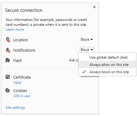
 Click it and Unblock the Notifications


